Brains on hormones
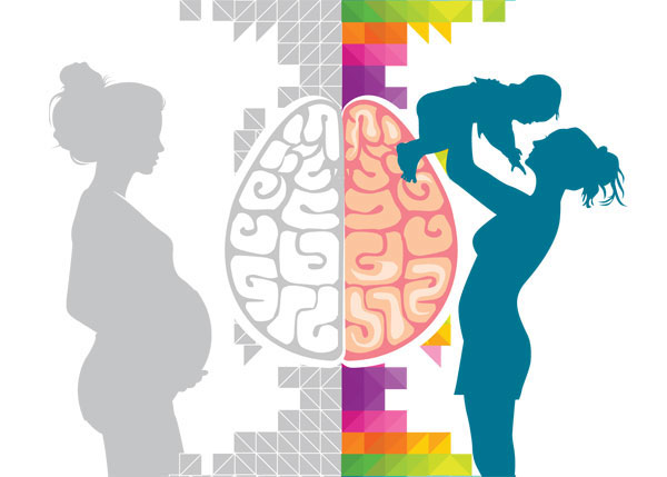
Many women mention to have memory lapses, cognitive impairment and difficulties to focus associated to pregnancy and motherhood. It is called «baby brain», «brain pregnancy» or «momnesia». In surveys, up to four-fifths of pregnant women report slight mental troubles such as problems remembering phone numbers or stringing a complex sentence together. It is considered a ‘myth’, but there are different evidences suggesting that the brain of pregnant women and mums with little children have structural and functional changes. One study found poorer short-term memory compared to other woman of the same age. In fact they found their memory to be similar to those in their sixties! It appears to alter exclusively new and complex memory tasks whereas regular, well-practiced habilities were not affected1. Another important evidence is that several studies have found that the brain decreases in size during pregnancy and increases in size after delivery. The changes follow a consistent time course in each woman. The reduction was maximal at term and returned to normal values by six months after delivery.2 The ventricular size showed a corresponding increase in size during pregnancy and a decrease in size after delivery.
The cause? Hormones! According to Louann Brizendine, director of the Women’s Mood and Hormone Clinic at the University of California, San Francisco, «There is 15 to 40 times more progesterone and estrogen marinating the brain during pregnancy» and « these hormones affect all kinds of neurons in the brain. By the time the woman delivers, there are huge surges of oxytocin that cause the uterus to contract and the body to produce milk — and they also affect the brain circuits.» 3 It is important not be too simplistic. Pregnancy also shuffles what get the attention of the mother and it is 100% normal to have memory lapses or be forgetful when she is busy, stressed, or short on sleep.

But if hormones change the brain, what about month cycle? The menstrual cycle is the regular natural change that occurs in the female reproductive system that makes pregnancy possible and it is governed by hormonal changes. The three sex steroid hormones, estrogen, progesterone and testosterone, vary cyclically and all functions that vary with the reproductive cycle can be considered to be mainly the result of gonadal steroid hormones.
A group of researchers from the Max Planck Institute for Human Cognitive and Brain Sciences has found that in parallel to the rhythm of estrogen across the menstrual cycle, the structure of the hippocampus varies 4. The research team did an exploratory, one-subject study. They acquired 30 longitudinal scans of a single, 32-year old, Caucasian female with a documented history of regular menstrual cycles (mean = 28 ± 1 day). The subject was physically healthy, with no history of any psychiatric, neurological, or chronic illnesses. The subject was right-handed, of normal weight (BMI 20.2), and free of medication, including no use of contraceptives or antidepressants. She was a non-smoker, with no history of alcohol use, drug abuse, pregnancy, or period of breast-feeding. Hormone assays for LH, estradiol, and progesterone were done and MRI images were acquired.
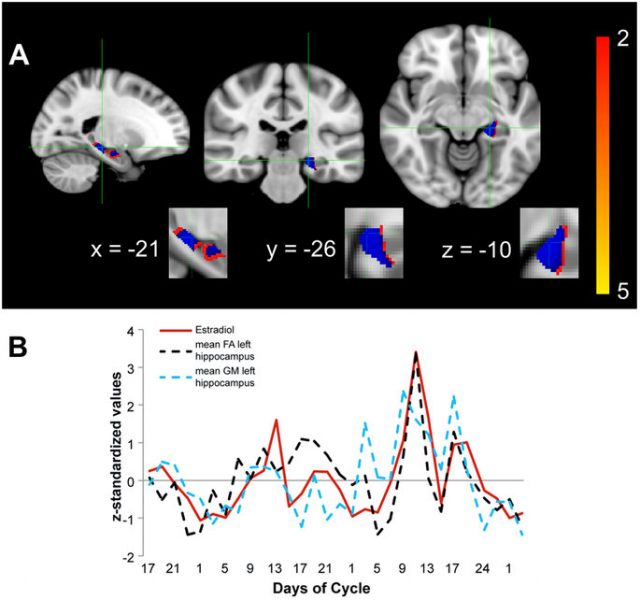
The authors found a significant positive correlation between fractional anisotropy values —a measure sensitive to changes in microstructural integrity— and estrogen in the hippocampus bilaterally, revealing a peak in fractional anisotropy closely paralleling ovulation. In parallel to the rising estrogen levels leading up to ovulation, there was an increase in the volume of the hippocampus, particularly in the grey-white matter boundary.
The hippocampus is located in the temporal lobe and plays a crucial role in our memories, our mood, and our emotions although it cannot be excluded that changes in vasculature contribute to the changes observed in diffusion scalar measures. Hippocampal abnormalities have been observed in several neuropsychiatric pathologies with prominent sexual dimorphism. The German study, a heavily sampled longitudinal dataset with detailed information on hormonal levels, could be a useful means to detect subtle brain changes related to the menstrual cycle and hormonal influence.
Understanding the effects of hormones in the brain is crucial for treating the premenstrual dysphoric disorder, a disorder that affects one in twelve women and is characterized by severe physical and psychological symptoms such as listlessness or mood swings. It is also promissory for understanding postnatal depression and even factors that are involved in healthy neuronal aging in all women. Up to know the information is contradictory 5. One study found a correlation between the number of children a woman had and her Alzheimer’s risk whereas another work suggested that breastfeeding and pregnancy may be protection factors against this neurodegenerative disease.
References
- Rendell PG, Henry JD (2008) Prospective-memory functioning is affected during pregnancy and postpartum. J Clin Exp Neuropsychol. 30(8): 913-919. ↩
- Oatridge A, Holdcroft A, Saeed N, Hajnal JV, Puri BK, Fusi L, Bydder GM (2002) Change in brain size during and after pregnancy: study in healthy women and women with preeclampsia. AJNR Am J Neuroradiol 23(1): 19-26. ↩
- Mann D Pregnancy Brain: Myth or Reality? WebMD ↩
- Barth C, Steele CJ, Mueller K, Rekkas VP, Arélin K, Pampel A, Burmann I, Kratzsch J, Villringer A, Sacher J (2016) In-vivo Dynamics of the Human Hippocampus across the Menstrual Cycle. Sci Rep 6: 32833. ↩
- Young E (2016) The real baby brain. New Scientist 3055: 36-39. ↩