A new imaging technique allows visualisation inside intact animals
A new imaging technique allows visualisation inside intact animals
One of the biggest challenges of medical imaging technologies is actually resolving the structures of interest, be it a tumour, a lung or a blood vessel, without the “noise” from other bodily parts like skin or muscle. A new imaging technique allows for visualisation inside intact animals at higher resolution than ever before.
A team of scientists from various institutions in Munich (Germany) has announced the development of wildDISCO 1, a new scanning technique based on previously used methods that allows for high resolution imaging of bodily structures labeled with antibodies. They tested their method on mice, dead mice, to be more precise as their process involves certain treatments which are not (yet) applicable to living tissue.
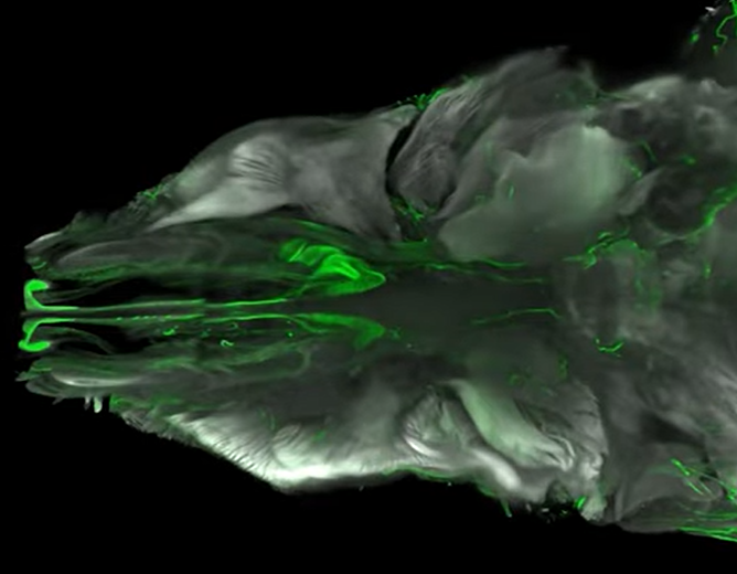
Their process involves using a cocktail of chemicals to remove fat and pigment from animal tissues, making them transparent. Next, the body is flooded with fluorescent antibodies, which label certain bodily regions like blood vessels or the brain. Then, the signals produced by these antibodies are picked up by scanners and stitched together into a three-dimensional, high-resolution map of the animal’s body.
https://www.youtube.com/watch?v=O38gTdZhUGk
Credit: Ertürk et al., 2023
This revolutionary technique has manyfold applications in research, from basic research on organ function to clinical research on drug development for diseases such as cancer, as it allows observing both the target and the effects of the treatment. It could also serve for improved diagnosis and follow-up, as its high resolution allows detecting even small lumps of tumour cells that would go undetected with current imaging technologies such as PET or MRI.
However, this revolutionary imaging technique that allows for visualisation inside animal bodies like never before is still far from being applicable to humans, at least living ones. In the future, the researchers that developed the technique expect to further implement the technique to expand its applications.
References
- Mai, H., Luo, J., Hoeher, L. et al. Whole-body cellular mapping in mouse using standard IgG antibodies. Nat Biotechnol (2023). doi: 10.1038/s41587-023-01846-0 ↩