Next generation nanoprobes for the microspectroscopic study of biosystems
Next generation nanoprobes for the microspectroscopic study of biosystems
Nonlinear processes are attractive in microscopy and spectroscopy since they can be excited with light in the near‐infrared, which offers several advantages, from a deep tissue penetration capability or a reduced photodamage due to the lower photon energy, to a spatially more confined probed volume, which can result in an improved lateral resolution. These processes include two‐photon‐excited fluorescence, second harmonic generation, stimulated Raman scattering and coherent anti‐Stokes Raman scattering microscopies, and imaging based on spontaneous hyper Raman scattering.
When multiphoton‐excited processes are complemented with their one‐photon‐excited counterparts, an unprecedented structural information from a sample can be obtained. Nonlinear processes, therefore, can be used for the visualization and monitoring of bioorganic molecules and structures, in particular for the investigation of basic chemical and physical processes in biosystems.
The nanoprobe used is a key factor. Actually, nanoprobes that could deliver chemical information through a vibrational signature with the requirements of fast nonlinear imaging would be extremely useful in the study of biological samples, as they allow truly multimodal optical imaging based on both chemical structure and morphology at the microscopic level. Several generations of optical nanoprobes and labels, strongly varying in chemical composition and size range, have been developed to match the specific requirements of multiphoton‐excited methods regarding functionality.

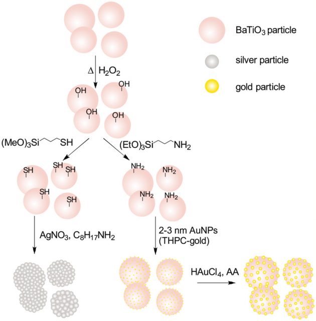
Now, a team of researchers reports 1 about the synthesis and optical characterization of gold‐ and silver‐coated barium titanate composite nanoparticles and demonstrate that they can be utilized as multifunctional optical nanoprobes for second harmonic generation, one‐photon surface enhanced Raman scattering, and surface enhanced hyper Raman scattering microscopies, thereby providing complementary information for imaging as well as for vibrational spectroscopy.
The researchers chose barium titanate as the core material because of its nonlinear optical properties and well established use as harmonic probe in second harmonic generation biomicroscopy. Surrounding the barium titanate nanocrystals with a shell of gold and silver nanoparticles allows taking advantage of the strongly enhanced Raman and hyper Raman processes in the near fields of these plasmonic structures, and thus enables sensitive vibrational probing of the local surface environment, which can provide complementary chemical and structural information of target samples.
The team shows that these composite nanoparticles can enhance Raman and hyper Raman scattering, while simultaneously acting as suitable second harmonic generation nanoprobes. This is demonstrated by applying them for combined second harmonic generation and vibrational imaging in macrophage cells. In order to understand how the optical properties of dielectric barium titanate particles are altered by the presence of the plasmonic shell, the researchers compare the experimental second harmonic generation data with theoretical simulations.
The theoretical models show that the optical properties of the BaTiO3 dielectric core depend on probing frequency, shape, size, and plasmonic properties of the surrounding gold nanoparticles so that they can be optimized for a particular type of experiment. These versatile, tunable probes give new opportunities for combined multiphoton probing of morphological structure and chemical properties of biosystems.
Author: César Tomé López is a science writer and the editor of Mapping Ignorance
Disclaimer: Parts of this article are copied verbatim or almost verbatim from the referenced research paper.
References
- Madzharova, F., Nodar, Á., Živanović, V., Huang, M. R. S., Koch, C. T., Esteban, R., Aizpurua, J., Kneipp, J. (2019) Gold‐ and Silver‐Coated Barium Titanate Nanocomposites as Probes for Two‐Photon Multimodal Microspectroscopy. Adv. Funct. Mater. 2019, 29, 1904289. doi: 10.1002/adfm.201904289 ↩