Platelets participate in hippocampal neurogenesis and cognitive function
Platelets participate in hippocampal neurogenesis and cognitive function
Platelets
Author: José R. Pineda got his Ph.D. from University of Barcelona in 2006. Since 2007 he has worked for Institut Curie and The French Alternative Energies and Atomic Energy Commission. Currently he is a researcher of the UPV/EHU. He investigates the role of stem cells in physiologic and pathologic conditions.
There is currently a consensus that promoting or engaging in physical activity produces tangible improvements in quality of life during aging. Experiments with aged mice that received blood plasma from exercising mice showed an enhanced cognition and improved neurogenesis demonstrating that the beneficial effects of exercise can be transferred 1. However, the molecular mechanisms underlying the beneficial effect of exercise on brain health remain poorly understood.
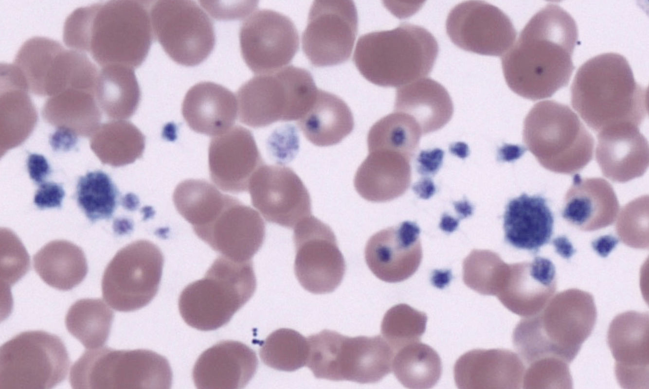
Platelets or also known as thrombocytes are fragments of cytoplasm derived from the megakaryocytes (bone marrow cells) that have no cell nucleus 2. It is known that platelets are a component of blood whose function is to react to bleeding from blood vessel injury by clumping, thereby initiating a blood clot. In the present work, Odette Leiter and collaborators published in Nature Communications journal an astonishing discovery: hippocampal neurogenesis can be rejuvenated improving cognitive function in the aged brain thanks to the secretion of some platelet-derived factors 3.
Platelets can be activated by exercise, and this is crucial to observe the changes for the exercise-induced increase in hippocampal precursor cell proliferation in aged mice. Platelet activation produces the secretion of what is called “Exerkines”. Exerkines are signaling moieties released in response to acute and/or chronic exercise that exerts their effects through endocrine pathways 4. Leiter and collaborators found that exerkine CXCL4/platelet factor 4 (PF4) was enough to reverse age-related regenerative and cognitive impairments in a hippocampal neurogenesis-dependent manner. However, how was this discovery possible? Let us see it step by step…
Activated platelets by acute exercise release humoral factors including the chemokine platelet factor 4 (PF4). It was already known that systemic PF4 is able to promote adult neurogenesis in young mice. They mimicked the increase in plasma PF4 levels by injecting 500 ng of PF4 into the tail vein of young mice every two day for one entire week. When sacrificed the animal they counted the number of immature neurons (doublecortin (DCX) positive cells) in a neurogenic region of the brain known as “dentate gyrus” (DG). They found that the increase of DCX+ cells was not due to the division of the stem cells, contrary to what is known after physical exercise (where is reported an increase of the nuclear proliferation marker “Ki67” from quiescent stem cells activating its proliferation). To confirm that systemic PF4 does not affect the recruitment of quiescent stem cells, they used a dual labelling paradigm in which when a cell divides (and to do so, the cell duplicate its genetic material) the analogues of thymidine CIdU and IdU are incorporated into the DNA labelling cell nuclei with both colors. Thus, administering CIdU (green) before PF4 treatment and IdU (red) after PF4 treatment they were able identify possible changes in the proportion of labelled cells respect to control (saline solution instead of PF4). However, they did not found any differences, demonstrating that previously quiescent cells were not being recruited into the proliferative cell cycle. Furthermore, they did not observe changes in the percentage of CIdU+ cells with or without IdU label, suggesting that the PF4 treatment did not affect cells that were proliferative prior to treatment. Their findings suggested that the treatment enhanced the neurogenic process at later stages through the survival or maturation of newborn neurons.
Next, they moved to a more variable-controlled experiments focusing in cell culture of neural stem and precursor cells to decipher its direct effects on cell proliferation, differentiation and cell morphology. Therefore, they were able to demonstrate that PF4 was able to be taken-up and internalized by the adult neural stem cells and did not stimulate cell proliferation (corroborating the observed results in vivo). Interestingly, they observed that a higher proportion of cells entered the G1/G0 cell cycle phases after PF4 treatment (that means: cells left their proliferative state) and started to differentiate into neurons. To check cell differentiation they did immunofluorescence against GFAP (astrocyte marker) and β-III-tubulin (immature neurons) observing a significant increase of β-III-tubulin+ cells in the PF4-treated cultures.
To check whether PF4 was required for the maintenance of hippocampal neurogenesis, they analyzed two neurogenic regions of adult brain, the subventricular zone (SVZ) and the (above-mentioned) DG in PF4 knockout mice (animals that do not express PF4). Surprisingly, they observed lower cell proliferation (Ki67+) and fewer DCX+ immature neurons in the DG but no changes in the SVZ, suggesting a specific effect of PF4 on adult hippocampal neurogenesis. They stimulated animals to do exercise housing them in cages with running wheels for 10 days. Control animals (with normal levels of PF4) increased neurogenesis meanwhile PF4 knockout mice cell proliferation was absent.
To understand which cell signaling pathways could trigger PF4 they did RNA sequencing and gene ontology, corroborating an upregulation of genes involved in neuronal differentiation, learning, memory and synaptic transmission. However, protein level analysis using western blot molecular biology technique did not reveal significant differences in presynaptic (synaptophysin) and postsynaptic (PSD-95) proteins. These results suggested that PF4 did not affect the synaptic composition. Morphological 3D-Sholl analysis is used to quantify neurite and cell projections complexity and again no changes were observed. Therefore, the researchers focused the study in the platelet proteome using a mass spectrometry-based proteomic analysis and found that exercise strongly altered it in both young and aged mice. Interestingly, selenoproteins and glutaredoxins (proteins with antioxidant capacity), as well as progranulin and properdin (proteins involved in anti-inflammatory and immunomodulatory activity) were upregulated in the platelets of young and aged mice. They did gene ontology in platelets of both young and aged mice (with or without exercise) to link to exercise-induced neurogenesis regulation. They collected blood from exercising and standard-housed aged mice and by flow cytometry observed activated platelets and they corroborated its effects neutralizing them using antiplatelet serum observing no neurogenic response. These astonishing results demonstrated, for the first time, the regulatory role of platelets in exercise-induced adult neurogenesis in aged animals.
Next, they focused the study on elderly mice (that is 20-month-old mice. Take into account that the lifespan of mice is about 24 months!). It was at this late stage in life that researchers observed notable changes on cognitive function induced by PF4 treatments. To do so, they used novel object location test and contextual fear conditioning behavioral tasks. They observed that PF4-treated animals displayed an improved recognition of the novel location, more frequent visits to the moved object suggesting an improvement in long-term memory capacity. Respect to fear conditioning, mice displayed a higher freezing score compared to control mice. Altogether, these results revealed that systemic PF4 treatment was able to recapitulate the beneficial effects of exercise by rejuvenating hippocampal neurogenesis and restoring cognitive function in the aged brain.
Finally, as all the results focused on the effects of PF4 on the survival and maturation of newborn neurons (DCX+ cells), a (big) unresolved question remained: what happens to the rejuvenating and neurocognitive effects in this paradigm if we eradicate the populations of (DCX+) immature neurons? Leiter and collaborators addressed this question administering PF4 to transgenic mouse “DCXDTR” (these mice express diphtheria toxin receptor (DTR) on the surface of DCX+ cells, so diphtheria toxin administration selective kills all newborn neurons) or PF4 and diphtheria toxin. Animals treated with diphtheria toxin reduced DCX+ cells and the cognition-enhancing effects of PF4 were absent, highlighting the necessity of neurogenesis for the PF4-mediated cognitive enhancement.
The work of Leiter and collaborators shows for the first time the effects of platelet activation response to exercise, identifying PF4 as factor that rejuvenates neurogenesis and cognition in the aged brain. These findings are the best example of systemic injection of exerkines as new promising factors for future therapeutics.
References
- Alana M Horowitz, Xuelai Fan, Gregor Bieri, Lucas K Smith, Cesar I Sanchez-Diaz, Adam B Schroer, Geraldine Gontier, Kaitlin B Casaletto, Joel H Kramer, Katherine E Williams, Saul A Villeda. “Blood factors transfer beneficial effects of exercise on neurogenesis and cognition to the aged brain”. Science. 2020 Jul 10;369(6500):167-173. doi: 10.1126/science.aaw2622. ↩
- Machlus KR, Thon JN, Italiano JE (April 2014). “Interpreting the developmental dance of the megakaryocyte: a review of the cellular and molecular processes mediating platelet formation”. British Journal of Haematology. 165 (2): 227–236. doi:10.1111/bjh.12758 ↩
- Odette Leiter, David Brici, Stephen J Fletcher, Xuan Ling Hilary Yong, Jocelyn Widagdo, Nicholas Matigian, Adam B Schroer, Gregor Bieri, Daniel G Blackmore, Perry F Bartlett, Victor Anggono, Saul A Villeda, Tara L Walker. “Platelet-derived exerkine CXCL4/platelet factor 4 rejuvenates hippocampal neurogenesis and restores cognitive function in aged mice” Nature Communications. 2023 Aug 16;14(1):4375. doi: 10.1038/s41467-023-39873-9. ↩
- Lisa S Chow, Robert E Gerszten, Joan M Taylor, Bente K Pedersen, Henriette van Praag, Scott Trappe, Mark A Febbraio, Zorina S Galis, Yunling Gao, Jacob M Haus, Ian R Lanza, Carl J Lavie, Chih-Hao Lee, Alejandro Lucia, Cedric Moro, Ambarish Pandey, Jeremy M Robbins, Kristin I Stanford, Alice E Thackray, Saul Villeda, Matthew J Watt, Ashley Xia, Juleen R Zierath, Bret H Goodpaster, Michael P Snyder. “Exerkines in health, resilience and disease.” Nat Rev Endocrinol. 2022 May;18(5):273-289. doi: 10.1038/s41574-022-00641-2. Epub 2022 Mar 18. ↩