Epigenetic reactivation of a tumor suppressor program in AML
Epigenetic reactivation of a tumor suppressor program in AML
Author: Marta Irigoyen is a postdoctoral researcher at CIC bioGUNE
Inactivation of tumor suppressor genes (TSGs) confers a cellular fitness advantage to human cancer cells, including in acute myeloid leukemia (AML), where it disrupts myeloid differentiation and promotes leukemogenesis. Loss-of-function mutations in TSGs are frequently observed in patients with hematologic malignancies 1, driving disease progression by blocking differentiation and enhancing self-renewal. Therefore, restoration of tumor suppressor activity represents a promising therapeutic strategy 2. Indeed, previous studies indicate that the therapeutic potential of this approach lies in the reversibility of TSG silencing 3. In the present work, Arnuk A and coworkers identified the transcriptional repressor ZBTB7A as a TSG that is downregulated in AML, especially within leukemia stem cells (LSCs). Its loss impairs differentiation and acceleraties disease progression through aberrant activation of inflammatory pathways 4. Importantly, the authors define an epigenetic strategy to therapeutically restore the ZBTB7A tumor suppressor function, demonstrating potent anti-leukemic activity in hematologic malignancies.
Firstly, to identify genes that suppress leukemia differentiation, the authors performed phenotypic CRISPR-Cas9 screens in two Cas9-expressing human AML cell lines. Differentiation was monitored via CD11b surface expression, detected using a fluorescently conjugated anti-CD11b antibody to identify AML blasts undergoing myeloid differentiation. Then, individual single guide RNA (sgRNA) abundance was analyzed by deep sequencing. In parallel, they conducted a positive-selection screen to identify genes whose loss promoted proliferation under differentiation-inducing conditions. Among the top-ranking genes identified was ZBTB7A, which encodes a POK domain that functions as a transcriptional repressor. To confirm their screen results, they individually transduced ZBTB7A sgRNAs (linked to a GFP reporter) into Cas9-expressing human AML cell lines and treated them with the differentiation-inducing agents. Results showed that ZBTB7A knockout (KO) cells exhibited reduced expression of the myeloid marker CD11b, and competition assays revealed an increased proportion of GFP+ cells in ZBTB7A KO versus control, indicating impaired differentiation. To examine Zbtb7a function in vivo, they used a Cre-inducible Cas9 mouse model and a Cre-expressing sgRNA vector encoding Zbtb7a sgRNA (which was transduced into hematopoietic stem and progenitor cells (HSPCs)) and transplanted into lethally irradiated host mice. Six weeks post-transplantation, they observed that Zbtb7a suppression led to a block in lineage-restricted progenitors and an expansion of HSC/MPP populations indicating that ZBTB7A loss suppresses AML differentiation. Conversely, cDNA of ZBTB7A (containing a GFP reporter) overexpression inhibited proliferation, promoted terminal differentiation and apoptosis, reduced colony formation, delayed leukemia progression, and prolonged survival of leukemia-bearing mice.
Mechanistically, chromatin immunoprecipitation sequencing (ChIP-seq) revealed that ZBTB7A binds to gene promoters and intronic regions. RNA sequencing (RNA-seq) analysis in Cas9-expressing AML cells showed transcriptional changes upon ZBTB7A loss. As expected for a transcriptional repressor, ZBTB7A KO led to widespread gene depression. Gene ontology (GO) analysis of the top upregulated pathways revealed key inflammatory responses and inflammatory cytokines and chemokines. Given their roles in shaping the inflammatory milieu of myeloid malignancies, they quantified TNF, IL-1β, MPO, and MMP-9 protein levels by enzyme-linked immunosorbent assay (ELISA) in conditioned media from ZBTB7A KO versus control AML cells. Consistent with transcriptomic findings, ZBTB7A KO significantly increased secretion of these inflammatory proteins. Conversely, ectopic overexpression of ZBTB7A led to global transcriptional repression. According with the previous data, the most significantly downregulated pathways upon ZBTB7A overexpression included inflammatory pathways. Notably, ZBTB7A loss resulted in increased expression of TNF and TNFAIP3, while its overexpression repressed these genes. Besides, ChIP-seq confirmed ZBTB7A binding at TNF and TNFAIP3 promoters.
To address the clinical relevance of their findings, they analyzed ZBTB7A expression using previously published single-cell RNA-seq (scRNA-seq) data of bone marrow specimens from diagnosed adult and pediatric AML patients 5, along with healthy donors. Hematopoietic stem cells (HSCs) from AML patients exhibited the lowest ZBTB7A expression, which progressively increased during myeloid maturation, linking expression to differentiation state. Analysis of an independent scRNA-seq cohort of patients 6 confirmed this pattern, showing the lowest ZBTB7A levels in HSC and early progenitor populations relative to mature myeloid cells. Given that high relapse rates and poor clinical outcomes in AML are largely driven by Leukemia Stem Cells (LSCs) 7, they examined the relationship between ZBTB7A expression and LSCs across three large AML cohorts 8910. Interestingly, ZBTB7A ranked among the top 2% of genes with strongest negative correlation to LSC17, indicating reduced expression is linked to stem-like states. Finally, analysis of AML patients from a different cohort 11 showed that lower ZBTB7A levels correlated with significantly worse overall survival. To explore the potential of reactivating ZBTB7A expression in AML, they developed a CRISPR-based screening approach to identify upstream regulators of TSGs. They designed fluorescent in situ hybridization (FISH) probes conjugated with a fluorophore (Alexa Fluor-647) to target the coding region of the ZBTB7A transcript. Next, they transduced AML Cas9 cells with a human genome-wide CRISPR/Cas9 knockout library 12. Ten days post-transduction, AML cells were fixed and incubated with the ZBTB7A FISH probe and sorted based on ZBTB7A expression levels. Deep sequencing was then performed and upstream transcriptional and epigenetic regulators whose ablation increased ZBTB7A expression in AML cells were prioritized. Among the top candidates KDM4B was identified and, interestingly, ENCODE ChIP-seq analysis showed that KDM4A and KDM4B histone demethylases were bound to the ZBTB7A promoter.
To restore ZBTB7A activity they used a pharmacological approach through KDM4 inhibition using QC6352 inhibitor (QC6352 is a potent pan-KDM4 inhibitor which effectively targeted KDM4A-C) 13. Treatment of human AML cell lines with QC6352 led to an approximately 3-fold increase in ZBTB7A mRNA levels. Moreover, QC6352 also induced a dose-dependent increase in ZBTB7A protein levels. Then, they conducted a time-course experiment and treated AML line cells with QC6352 and found that induced myeloid differentiation followed by apoptosis. Moreover, KDM4 inhibition induced robust anti-leukemic activity and terminal differentiation across a broad range of AML cell lines and PDX models.
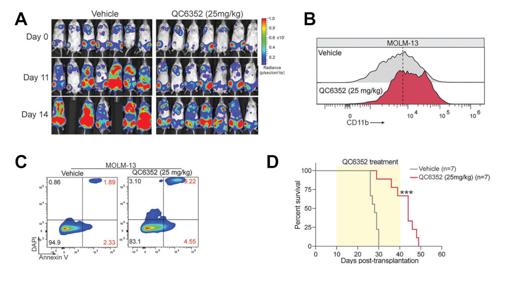
To assess the in vivo efficacy of KDM4 inhibition, they tested QC6352 in both AML cell line and PDX models. AML cells expressing firefly luciferase (an enzyme that emits photons after degradation of luciferine compound) were transplanted into immune-deficient recipient mice. Upon disease establishment, mice were treated with QC6352. Notably, bioluminescent (photon) quantification and imaging of leukemia-bearing animals treated with QC6352 showed marked delay in AML burden. Consistent with in vitro results, QC6352 induced myeloid differentiation and apoptosis in vivo at day 10, reduced AML burden, and significantly extended survival. They further extended these observations in two AML PDX models. Following engraftment in NSG-SGM3 mice, they were treated with QC6352 or vehicle. Remarkably, QC6352 treatment significantly reduced human AML cells in blood and bone marrow and decreased spleen infiltration and weight. They also observed that QC6352 treatment increased myeloid differentiation and apoptosis and significantly prolonged the survival of leukemia-bearing PDX mice compared to vehicle controls.
In summary, this study identifies the transcriptional repressor ZBTB7A as a TSG downregulated in AML, associated with poor survival outcomes. To uncover therapeutic strategies, they developed a methodology pinpointing the KDM4 family of histone demethylases as a vulnerability to restore ZBTB7A function. Thus, pharmacological inhibition of KDM4 upregulated ZBTB7A expression, promoted terminal differentiation of leukemic cells and prolonged mice survival. These findings reveal critical regulatory mechanisms of ZBTB7A and support epigenetic therapy as a promising strategy to reactivate its tumor suppressor function in hematologic cancers.
References
- Papaemmanuil E, Gerstung M, Bullinger L, Gaidzik VI, Paschka P, Roberts ND, Potter NE, Heuser M et al. Genomic Classification and Prognosis in Acute Myeloid Leukemia. N Engl J Med. 2016; 374: 2209-2221. PMID: 27276561 DOI: 10.1056/NEJMoa1516192. ↩
- Ventura A, Kirsch DG, McLaughlin ME, Tuveson DA, Grimm J, Lintault L, Newman J, Reczek EE, Weissleder R et al. Restoration of p53 function leads to tumour regression in vivo. Nature. 2007; 445: 661-665. PMID: 17251932 DOI: 10.1038/nature05541. ↩
- Will B, Vogler TO, Narayanagari S, Bartholdy B, Todorova TI, da Silva Ferreira M, Chen J, Yu Y, Mayer J, Barreyro L et al. Minimal PU.1 reduction induces a preleukemic state and promotes development of acute myeloid leukemia. Nat Med. 2015; 21: 1172-1181. PMID: 26343801 DOI: 10.1038/nm.3936. ↩
- Arnuk A, Han C, Lawal AE, Wang B, Karma S, Zhang Z, Yassouf MY, Rajendran SH et al. (2026) Epigenetic reactivation of the tumor suppressor ZBTB7A by KDM4 inhibition in human acute myeloid leukemia. Sci Transl Med. doi: 10.1126/scitranslmed.ady2936. ↩
- Lasry A, Nadorp B, Fornerod M, Nicolet D, Wu H, Walker CJ, Sun Z, Witkowski MT et al. An inflammatory state remodels the immune microenvironment and improves risk stratification in acute myeloid leukemia. Nat Cancer. 2022; 4: 27-42. PMID: 36581735 DOI: 10.1038/s43018-022-00480-0. ↩
- Zeng AGX, Iacobucci I, Shah S, Mitchell A, Wong G, Bansal S, Chen D, Gao Q et al. Single-cell Transcriptional Atlas of Human Hematopoiesis Reveals Genetic and Hierarchy-Based Determinants of Aberrant AML Differentiation. Blood Cancer Discov. 2025; 6: 307-324. PMID: 40294241 DOI: 10.1158/2643-3230.BCD-24-0342. ↩
- Ng SW, Mitchell A, Kennedy JA, Chen WC, McLeod J, Ibrahimova N, Arruda A, Popescu A et al. 17-gene stemness score for rapid determination of risk in acute leukaemia. Nature. 2016; 540: 433-437. PMID: 27926740 DOI: 10.1038/nature20598. ↩
- N. Cancer Genome Atlas Research, Ley TJ, Miller C, Ding L, Raphael BJ, Mungall AJ, Robertson A, Hoadley K et al. N Engl J Med. 2013; 368: 2059-2074. PMID: 23634996 DOI: 10.1056/NEJMoa1301689. ↩
- Tyner JW, Tognon CE, Bottomly D, Wilmot B, Kurtz SE, Savage SL, Long N, Schultz AR et al. Functional genomic landscape of acute myeloid leukaemia. Nature. 2018; 562: 526-531. PMID: 30333627 DOI: 10.1038/s41586-018-0623-z. ↩
- Abbas HA, Mohanty V, Wang R, Huang Y, Liang S, Wang F, Zhang J, Qiu Y et al. Decoupling Lineage-Associated Genes in Acute Myeloid Leukemia Reveals Inflammatory and Metabolic Signatures Associated With Outcomes. Front Oncol. 2021; 11: 705627. PMID: 34422660 DOI: 10.3389/fonc.2021.705627 ↩
- Mitschka S, Mayr C, Endogenous p53 expression in human and mouse is not regulated by its 3’UTR. Elife. 2021; 10: e65700. PMID: 33955355 DOI: 10.7554/eLife.65700. ↩
- Doench JG, Fusi N, Sullender M, Hegde M, Vaimberg EW, Donovan KF, Smith I, Tothova Z et al. Optimized sgRNA design to maximize activity and minimize off-target effects of CRISPR-Cas9. Nat Biotechnol. 2016; 34: 184-191; PMID: 26780180 DOI: 10.1038/nbt.3437. ↩
- Chen YK, Bonaldi T, Cuomo A, Del Rosario JR, Hosfield DJ, Kanouni T, Kao SC, Lai C, Lobo NA, Matuszkiewicz J, McGeehan A, O’Connell SM, Shi L, Stafford JA, Stansfield RK, Veal JM, Weiss MS, Yuen NY, Wallace MB, Design of KDM4 Inhibitors with Antiproliferative Effects in Cancer Models. ACS Med Chem Lett. 2017, 8: 869-874. PMID: 28835804 DOI: 10.1021/acsmedchemlett.7b00220. ↩