Looking through opaque materials
A translucent material allows light to pass through, but if we try to look an object behind such a material, the image will appear blurred or distorted. Think, for example, of the semitransparent glasses commonly used as showers screens. The reason why the image deforms when passing through the glass is the scattering of light. The initial trajectory of the light is disturbed by the molecules in the glass, which scramble the spatial information, disabling in this way the formation of a sharp image. In situations where the scattering is very strong, such as for example a heavy-fog day, the visibility may be completely impaired. The scattering media behaves as an opaque material and objects become invisible.
Diffusion of light in media results sometimes in unwanted effects. For example, it may become a severe obstacle in diagnosis techniques. Nowadays, multiple disciplines, ranging from biosciences to nanotechnology, use optical imaging techniques as an essential tool for diagnosis. The so called optical coherence tomography is by now an established medical imaging technique used, for instance, to obtain images of the retina and coronary arteries. Another example of optical imaging application is the characterization of semiconductor wafers, such as surface and cross-section imaging. Optical coherence tomography can obtain sharp image through semitransparent media, but only for moderate scattering. For stronger scattering, its applicability is limited. Other methods are more successful with higher scattering but they are invasive, in the sense that they require being able to modify the conditions around the object (like placing a detector or a nonlinear material). The range of applications of these methods is bounded by both its invasive nature and its limited resolution capabilities.
Being able to obtain a sharp image when “looking” through a strong diffuser has become possible for a team from the MESA+ Institute for nanotechnology at the University of Twente in the Netherlands. The team, led by Dr. Allard Mosk, succeeded in imaging a fluorescent object hidden behind a scattering layer 1.
The method devised by the team of Mosk has a relatively simple setup, which is schematically shown in Figure 1. An opaque glass is placed between the object to be imaged and a detector. The test object used is the Greek letter “π“, about 50 μm across, written with fluorescent ink. The screen is a ground-glass diffuser. To excite the object fluorescence, a green diode laser is fired towards the object, however, by first crossing through the opaque screen. A speckle pattern emerges at the other side of the screen and this is superimposed on the object.
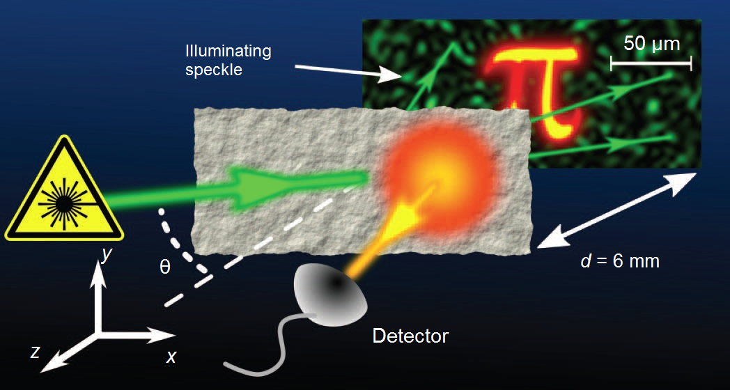
Speckle effect is a well-known phenomenon that results from the interference of many waves of the same frequency, with random phases and amplitudes. A familiar example is when shining a laser beam on a rough surface. A random pattern shows visible with dark and bright areas that resemble a surface with salt and pepper spread on top. In our setup described above, those areas of the object illuminated by the bright parts of the speckle will create more fluorescence that those areas illuminated by darker parts of the speckle. The total intensity reaching the detector is proportional to the integral of the speckle pattern over the entire area of the object. Despite the spatial information carried by the light is scrambled by the diffuser screen and the detector is not able to resolve the image, the transmitted fluorescence still retains the information of the overlap between the object response and the speckle intensity pattern on the object.
Very ingeniously, the team of Mosk devised a method to decode such information and extract the object fluorescence response, creating in this way the desired image. To achieve it, they scanned the angle of incidence of the green laser on the screen. This resulted in a speckle pattern moving across the object. The key element here is that when this rotation angle of the laser is sufficiently small, the speckle pattern does not change its spatial distribution but only translates over a distance. This is called “memory effect” and it allowed the group of researchers to mathematically separate object and speckle autocorrelations in the detected intensity. By scanning the laser angle within a small range, the intensity on the detector contains, for all the measurements within the range, the overlap between the object response and the very same speckle pattern.
After measuring the total intensity as a function of the scan angle, and using mathematical manipulation of the data, the team separated the autocorrelation of the speckle pattern from the autocorrelation of the fluorescent object. Formally, an autocorrelation cannot be inverted to form the original object, although, an approximate inversion is still possible. Iterative algorithms developed for astronomy and X-ray scattering applications were applied here to reconstruct the image. The result of the inversion shows the resemblance between the microscope image of the object obtained before placing the opaque screen and the image obtained after processing the detector signal.
To show the feasibility of the technique in biological systems, the authors applied the same method to image a slice of stem of lily of the valley, a plant that presents intracellular auto fluorescence. The obtained image showed again a faithful similarity with the image obtained by using conventional microscopy.
The method developed by Mosk and his colleagues offers a great potential for non-invasive imaging. By decreasing the size of the speckle spots, the resolution can be increased up to the diffraction limit. The authors also point to using the same concept but with other signals that also depend on the speckle intensity. This can allow, for example, sampling in a third dimension, to obtain three-dimensional images.
Both the potential applications of the technique and the simplicity of the experimental setup are big achievements of this work. In fact, the research was considered one of the breakthrough publications in 2012 by Nature. “Imaging through opaque materials” found its place among the top ten of the year, next to the discovery of Majorana fermions, the violation of time-reversal symmetry and neutrino-based communication. Certainly, it is expected that optical imaging techniques will experience in the coming years a great development as a result of this work.
References
- J. Bertolotti, E.G. van Putten, C. Blum, A. Lagendijk, W.L. Vos, A.P. Mosk, “Non-invasive imaging through opaque scattering layers“, Nature 491, 232 (2012) (DOI: 10.1038/nature11578) ↩