From bones to 3D(s): diffusion, didactics and diagnostics
From bones to 3D(s): diffusion, didactics and diagnostics
Author:
Dany Continho-Nogueira is manager of the PACEA-Transfert Unit of the University of Bordeaux
Methodological developments in medical imaging, computer sciences and rapid prototyping technologies offer new possibilities for research and formation in anthropology, archaeology and curation procedures but also for the benefit of the cultural heritage.
To conduct their studies on rare fossil specimens, anthropologists and archaeologists have to deal with two challenges. First, the specimens of interest are spread all around the world and then, it is not always possible to manipulate these fragile and precious remains because of conservation rules.
To overcome these obstacles, the use of the medical imaging progressively increased. A 3D digital chain, Vircopal® (Virtual Collection of paleospecimens)1, was developed by four researchers from two laboratories of Bordeaux University, PACEA (De la Préhistoire à l’Actuel : Culture, Environnement, Anthropologie) and LaBRI (Laboratoire Bordelais de Recherche en Informatique). This chain is based on three main points:
-
X-ray (micro-)Computed Tomography (the same technology used in medicine to scan patients);
-
Treatment of the medical images with the software TIVMI® and the HMH (half maximum height) algorithm 2 which accurately determines the interfaces between the different tissues 3;
-
High-quality 3D printing of the model.
This 3D digital chain produces faithful copies of the remains at different scales and provides access to their internal structures without damaging the pieces. Applications can cover a wide range, such as the preservation of the cultural heritage, research, education and reconstructive surgery.
The diffusion and preservation of the cultural Heritage
As previously stated, the fossils and the archaeological remains are not easily accessible to researchers because of their various locations and it is not always possible to manipulate them. Until the recent development of the medical imaging in anthropology and the 3D printing technology, the specimens were cast and then exchanged between research laboratories and/or museums. However, the actual conservation rules prohibit the casting of fragile archaeological remains.
Being non-invasive and non-destructive, the x-ray acquisition scan followed by the 3D reconstruction and printing of the resulting model provide a secure solution to faithfully replicate the remains, a solution that is also more precise than castings. The 3D data obtained can be stored and printed in multiple copies facilitating the preservation, dissemination and the enhancement of the cultural Heritage. These replicas can be exposed in museums and manipulated by the public, unlike many original pieces.
A didactic tool for the students
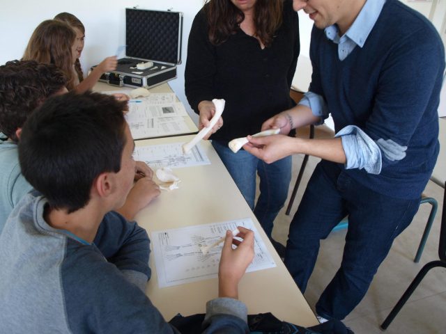
The universities collections rarely possess a wide variety of archaeological pieces and human remains. In anthropology, it is difficult to have access to bones affected by pathologies (syphilis, tuberculosis, rickets, etc.) or with a rare morphology (supernumerary bones or activity-related skeletal morphologies, for example). The reproduction of bones or archaeological remains presenting particular characters by 3D printing allows to enrich the collections and to provide pedagogical kits for the formation of the students in anthropology, palaeontology, archaeology and medicine.
Diagnoses paleopathologies and treat bones trauma
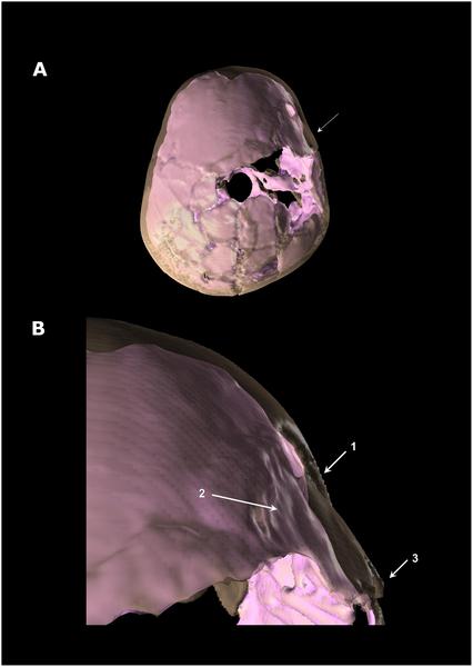
This technology is also a remarkable tool to study and to better understand the remains. The three-dimensional imaging methods can be used for different kinds of studies on bones but it is particularly interesting for paleopathology. The precision of this technology allows, for example with child’s bones, understanding the microarchitecture and the organisation of the different tissues4 but also to distinguish physiological growth processes from phenomena due to pathologies5, two results which could not be demonstrated with traditional studies or without the destruction of the bones. Using Vircopal® technology and the software TIVMI®, researchers could better explore the cranioencephalic trauma of the Qafzeh 11 child and its consequences on the development of his brain and skull. They demonstrated that the trauma caused significant neurological and psychological disorders; this is probably why this young individual received a ritual treatment different from the others burials found in Qafzeh6.
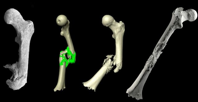
This technology is not only used to diagnose diseases on ancient remains, it has been successfully tested in reconstructive surgery. A soldier mandible shot by a bullet was 3D reconstructed by symmetry and 3D print. This model allowed the surgeon to shape a bone graft perfectly suited to the patient’s morphology and thus reduce the duration of the intervention to 3 hours.
With the financial assistance of the SATT Aquitaine (AST) and the Aquitaine Region, a technology transfer unit, PACEA-Transfert, was created to promote economically the Vircopal® process. This cell is attached to the research laboratory PACEA and is administratively managed by the ADERA (Association pour le Développement de l’Enseignement et des Recherches auprès des universités, des centres de Recherche et des entreprises d’Aquitaine).
References
- Coqueugniot H, Dutailly B, Desbarats P, Dutour O (2013) Procédé de modélisation d’une pièce formée de tissu osseux. Bordeaux and Aix-Marseille Universities, France. French patent n°1151284. ↩
- Spoor CF, Zonneveld FW, Macho GA (1993) Linear measurements of cortical bone and dental enamel by computed tomography: applications and problems. Am J Phys Anthrop 91: 469–484. doi: 10.1002/ajpa.1330910405 ↩
- Guyomarc’h P, Santos F, Dutailly B, Desbarats P, Bou C, et al. (2012) Three-dimensional computer-assisted craniometrics: a comparison of the uncertainty in measurement induced by surface reconstruction performed by two computer programmes. Forensic Sci Int 219: 221–227. doi: 10.1016/j.forsciint.2012.01.008 ↩
- Colombo A., Dutour O., Tillier A.-M. et Coqueugniot H. (2013). Asymétrie de la microarchitecture trabéculaire chez l’enfant : Analyse microscanner de l’extrémité proximale de l’humérus de sujets immatures de la collection ostéoarchéologique de Saint-Martin de Cognac (Charente, France).1838èmes Journées de la Société d’Anthropologie de Paris, Paris, 23-25 janvier 2013 ↩
- Rittemard Ch., Colombo A., Desbarats P., Dutailly B., Coqueugniot H. et Dutour O. (2014) Analyse macro- et micro-morphologique de l’os cortical en croissance : physiologie versus pathologie du périoste 1839èmes Journées de la Société d’Anthropologie de Paris, Montpellier, 28-31 janvier 2014 ↩
- Coqueugniot H, Dutour O, Arensburg B, Duday H, Vandermeersch B, et al. (2014) Earliest Cranio-Encephalic Trauma from the Levantine Middle Palaeolithic: 3D Reappraisal of the Qafzeh 11 Skull, Consequences of Pediatric Brain Damage on Individual Life Condition and Social Care. PLoS ONE 9(7): e102822. doi:10.1371/journal.pone.0102822 ↩
2 comments
Bonjour, tout a fait passionant pour l’education et les expositions : on sort des faux platres ! vous pourriez peut-etre faire l’imagerie 3D des squelettes de Cro-Magnon , et d ailleurs en profiter pour faire aussi leur analyse ADN qui n a toujours pas ete faite : liens familiaux ou pas ? mystere….
Cordialement
Francois Pelatan
(Desole pour les accents : clavier )
[…] Posted in Noticias, Science, Technology, Computer science, Materials, Medicine | 0 comments […]