A microscopy method to look at amyloid protein structure
A microscopy method to look at amyloid protein structure
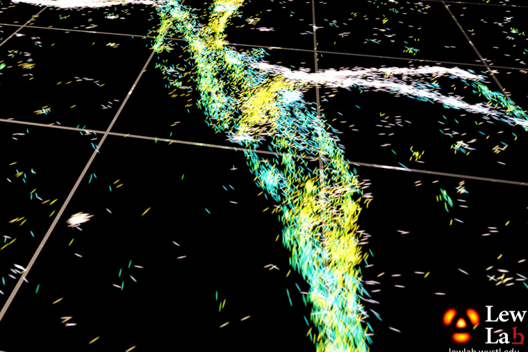
Amyloid plaques are a hallmark of some neurodegenerative diseases like Alzheimer’s or Parkinson’s. Even though the exact role of amyloid protein in the disease is not clear, being able to follow amyloid protein misfolding and plaque formation could be a step forward for the study of the disease. Now, Matthew Lew’s lab has developed 1 a new optical microscopy method that allows to look at the orientation of single molecules in the amyloid protein.
Since all biochemical reactions involve interactions among molecules, and those imply movement and certain displacement, it might be possible that these misfolded protein aggregates causes disruptions to these interactions, leading eventually to death. That is why the team developed this imaging methodology to be able to understand these molecular movements.
To get the most information of the technique, and determine orientation, the researchers used a fluorescent label directly attached to each single amyloid molecule, and measured extra parameters like light polarization. They followed over time the changes in orientation, the movement in the end, that every molecule experienced every time a new one joined an aggregate.
This microscopic method is open to imaging labs and can look into amyloid protein orientation
What’s most interesting about their imaging setup is that, according to them, everyone could have one, in that they used commercial components common to single-molecule super-resolution microscopy. In a commendable move towards Open Science, they even shared the analysis code used in the project.
By characterizing differences in the behaviour of these molecules, the researches want to get an insight into long-term amyloid aggregate formation, dispersion and its organization.
Their hope is that their technique can help understand the disease and eventually lead to potential treatments or even better ways of preventing amyloid disease.
References
- Tianben Ding, Tingting Wu, Hesam Mazidi, Oumeng Zhang, and Matthew D. Lew (2020) Single-molecule orientation localization microscopy for resolving structural heterogeneities between amyloid fibrils Optica doi: 10.1364/OPTICA.388157 ↩
1 comment
[…] Gaixotasun neurodegeneratiboetan ohikoak diren amiloide plaken irregulartasunak aztertzea ahalbidetzen du mikroskopia metodo berri batek. Ohiko piezekin egina eta kode irekia du gainera. Rosa García- Verdugoren A microscopy method to look at amyloid protein structure […]