Viruses at the edge of life

The definition of life is not a simple one. According to classical textbooks, “living things are born, grow, reproduce and die”. But one doesn’t have to look too far to find some caveats in this interpretation. For example, when bacteria divide, can we really say that one is “giving birth” to the other? The part about death is also controversial: there are organisms that have been around for thousands of years, like the Californian bristlecone pines, and there are even animals that don’t die.
Many definitions instead focus on the fact that living things are made of cells. Roughly speaking, cells are semi-isolated ‘containers’ that provide a favourable environment for specific chemical reactions to occur. Simple organisms, like bacteria or amoebas, are made of a single cell. Others, such as animals and plants, display higher levels or complexity, with cells organized in tissues and organs.
A definition of life based on its cellular nature has the asset of allowing to include in the same category beings as different as fungus and elephants. However, it leaves out an important group that has been troubling researchers for the last hundred years: viruses.
What’s a virus?
Viruses are neither dead nor alive, they sit on the edge of both worlds. Their structure is very simple, consisting solely of genetic material, be it DNA or RNA, surrounded by a protein structure called capsid.
Viruses have genetic material and they can replicate, two important traits that would argue in favour of qualifying them as living things. The problem is that normally viruses are metabolically inert. To become ‘alive’ they need to infect cells. Once inside, they hijack their machinery, create copies of themselves and leave in search of new victims.
Until recently, it was believed that all viruses were small. The average virus is about one one-hundredth the size of an average bacterium and can’t be seen using a regular light microscope. In fact, their size was what allowed their discovery, as they were initially identified as “infectious agents” small enough to pass through very narrow filters that retained bacteria.
This all changed in 2003, when an incredible finding forced virologists to forget everything they thought they knew about viruses.
Giant viruses
In 1992, following a pneumonia outbreak in a hospital in Bradford, England, water samples from a cooling tower were examined in search of the causing agent. The water contained free-living amoebas, unicellular organisms that could be causing the outbreak. But the researchers also noticed some strange parasites living inside the amoebas. Due to their size, they assumed they were bacteria. However, attempts to isolate them or amplify their DNA using standard bacterial techniques were unsuccessful.
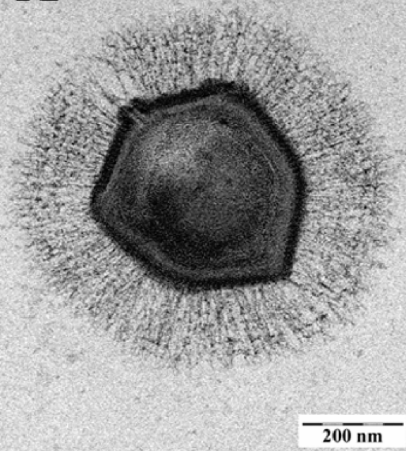
Years passed and in the early 2000s a group of researchers from the Université de la Mediterranée, in Marseille, used electronic microscopy to look at the strange parasite in more detail. To their surprise, they observed it had an icosahedral shape typically found in viral capsides. Further experiments confirmed that what they were looking at was not a bacterium, but rather a new form of giant virus. The results were published in Science1 and the new virus was called ‘Mimivirus’, for ‘mimicking microbe virus’.
Since then, there has been a very successful “virus rush” to search for other giant viruses using amoebas as bait. The approach has proven fruitful: since 2003, nine new families have been described. Giant viruses have been found in sewers, in seawater, in ponds, and, amazingly, in Siberian soil which has been kept frozen for 30,000 years.
The viruses that came in from the cold
The properties of the Siberian permafrost make it especially suited to look for long-term surviving micro-organisms. So researchers took a soil sample from 30 meters below the surface of a late Pleistocene sediment in the Kolyma region, and used it to infect amoebas in the lab. They found that the soil sample contained two types of giant viruses. Their findings were reported in two papers published in Proceedings of the National Academy of Sciences.
In 2014, when the first paper 2 came out, there were two clearly distinct groups of giant viruses, called ‘Megavirus’ and ‘Pandoravirus’. Megaviruses have a pseudo-icosahedral structure and replicate in the cytoplasm of the infected amoebas, while Pandoraviruses have a larger size, a longer genome, amphora-shaped capsids, and replicate in the nucleus.
The first virus looked a bit like both: it resembled Pandoravirus in shape and size, but its replication mechanism was similar to Megavirus. The researchers called it Pithovirus sibericum. Pithos is Greek for “large container” or “amphora”, and refers to Pandora’s box, the one that, according to the myth, contained all the evils in the world.
Each Pithovirus particle is about 1.5 μm, a very large size even for giant viruses standards. For comparison terms, that’s 50 times the size of the polio virus and 10 times the size of the HIV. But surprisingly, its genome is rather small. It has only 600,000 base pairs, about four times less than the Pandoravirus genome.
The second virus, described a year later3, was called Mollivirus sibericum, “molli” coming from a French word meaning ‘soft or flexible’. The new species also failed to fit any of the existing categories. It is quite small for a giant virus (0.6 μm, about three times smaller than the Pithovirus), but has a similar-sized genome. Like Pandoravirus, it replicates around the nucleus of the infected amoeba.
Both papers end with a word of caution. Global warming and mining activities along the northern coast of Siberia could have unpredictable effects on the permafrost. Giant viruses seem to infect specifically amoebas, but it’s not far-fetched to think that other dormant viruses could infect humans. As one of the authors put it: “What may be in those layers is very worrying”.
References
- A giant virus in amoebae. La Scola B et al. Science (2003)299:2033. doi: 10.1126/science.1081867 ↩
- Thirty-thousand-year-old distant relative of giant icosahedral DNA viruses with a pandoravirus morphology. Legendre M et al. Proc Natl Acad Sci U S A. (2014) 111:4274. doi: 10.1073/pnas.1320670111 ↩
- In-depth study of Mollivirus sibericum, a new 30,000-y-old giant virus infecting Acanthamoeba. Legendre M et al. Proc Natl Acad Sci U S A. (2015) 112:E5327. doi: 10.1073/pnas.1510795112 ↩