Secretome of dental pulp stem cells as a potential therapy for androgenetic alopecia
Secretome of dental pulp stem cells as a potential therapy for androgenetic alopecia
Author: José R. Pineda got his Ph.D. from University of Barcelona in 2006. Since 2007 he has worked for Institut Curie and The French Alternative Energies and Atomic Energy Commission. Currently he is a researcher of the EHU. He investigates the role of stem cells in physiologic and pathologic conditions.
Androgenetic alopecia is characterized by a progressive hair loss. This represents a cosmetic issue, treated with a limited set of pharmacological tools such as minoxidil or finasteride. While these therapies can be effective in some patients, their results are often inconsistent and frequently accompanied by unwanted side effects. This has driven researchers to search for alternative approaches that do not simply mask the problem, but instead target the biological processes underlying hair follicle degeneration. A recent study by Luo and colleagues 1 explores a particularly intriguing possibility: the use of tiny biological particles, known as exosomal extracelular vesicles, a tiny cargos that cells secrete to communicate and instric other cells. These are extremely small vesicles, essentially microscopic “packages” released by cells that contain proteins, signaling molecules, and genetic material. Their function is to communicate with other cells and influence their behavior. They plan to use this secretion to use its messages to stimulate hair regeneration through vascular and cellular repair mechanisms.
At the heart of this work lies a concept that may sound surprising at first. The researchers focused on dental pulp stem cells, a stem cell source obtained from the dental pulp of the third molars. These cells are known for their strong regenerative potential 234. Rather than grafting these cells directly, however, the study uses the exosomes that they naturally produce. Because exosomes can exert powerful effects without requiring direct cell transplantation, they are increasingly seen as promising tools for regenerative medicine. To understand how these exosomes might help with hair loss, the researchers designed a combination of laboratory and animal experiments. They first isolated dental pulp stem cells from human donors and confirmed that these cells retained their typical properties, including the ability to differentiate into multiple tissue types. From these cells, they extracted exosomes using a series of centrifugation steps designed to separate them from other cellular debris. The resulting particles were then carefully characterized using electron microscopy, which allowed the researchers to visualize their structure, and nanoparticle tracking analysis, which provided information about their size. These analyses confirmed that the isolated vesicles had the expected morphology and molecular markers of exosomes.
The next step was to determine whether these exosomes could influence the behavior of dermal papilla cells, a specialized cell population located at the base of hair follicles. These cells play a central role in regulating hair growth by producing signals that activate hair follicle stem cells. When dermal papilla cells lose functionality (as happens in androgenetic alopecia) hair follicles shrink and eventually stop producing visible hair. In cell culture experiments, the researchers exposed dermal papilla cells to the exosomes and observed their effects over time. Importantly, they used a well-established model of hair loss by treating the cells with dihydrotestosterone (DHT), a hormone known to impair hair growth and mimic the conditions of androgenetic alopecia. Under these conditions, dermal papilla cells typically show reduced proliferation and weakened regenerative capacity. However, when exosomes were added to the system, this negative effect was largely reversed. The treated cells proliferated more actively and showed improved migration, suggesting that they were regaining functionality. These functional improvements were accompanied by molecular changes. The researchers measured the expression of key genes associated with hair induction, such as alkaline phosphatase (ALP) and (alpha–Smooth Muscle Actin) α-SMA. They found that their levels increased significantly after exosome treatment. This indicates that the cells were not only surviving better, but were also recovering their ability to promote hair growth.
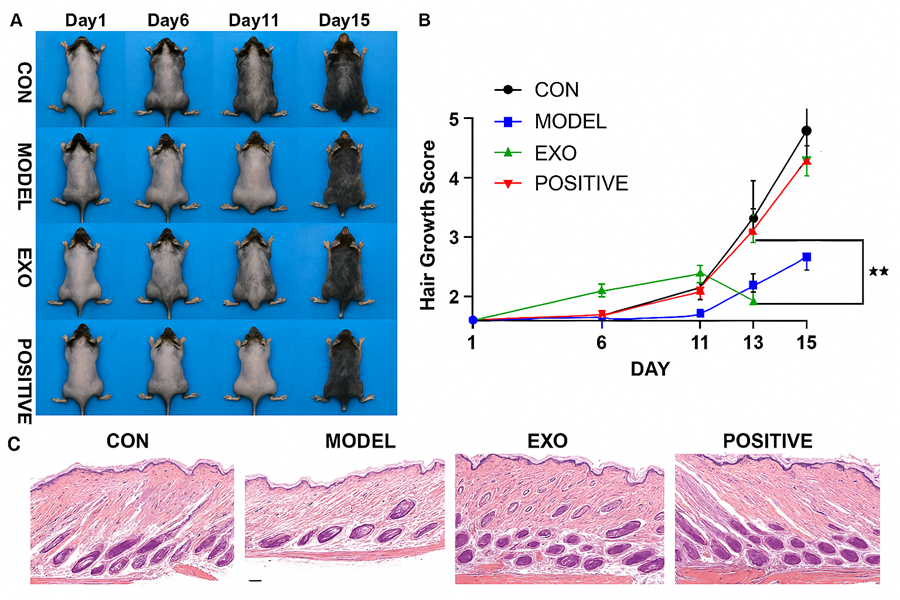
One of the most interesting aspects of the study lies in its attempt to uncover the biological mechanism behind these effects. To do this, the team used transcriptomic analysis (a method that allows scientists to examine changes in the activity of thousands of genes simultaneously). This approach revealed that exosome treatment activated a signaling pathway known as PI3K/Akt, which is widely involved in cell survival, proliferation, and tissue regeneration. At the same time, the treated cells showed increased expression of VEGFA (vascular endotelial growth factor A), a key molecule that promotes the formation of new blood vessels. This connection between cellular signaling and vascularization is crucial. Hair follicles are highly dynamic structures that require a constant supply of oxygen and nutrients to sustain growth. When blood flow around the follicle is compromised, the growth phase of the hair cycle is shortened, leading to progressive miniaturization of the follicle. By enhancing angiogenesis (the formation of new blood vessels) the exosomes appear to restore the microenvironment necessary for healthy hair growth. To confirm that this pathway was indeed responsible for the observed effects, the researchers performed inhibition experiments using LY-294002 a molecule that blocks PI3K/Akt activity. When this pathway was inhibited, the beneficial effects of the exosomes were almost completely abolished. The dermal papilla cells lost their ability to proliferate and to express hair-inductive markers, demonstrating that activation of PI3K/Akt signaling is not just associated with the process, but is required for it to occur. While these in vitro experiments provide valuable insights, the true test of any therapeutic approach lies in its performance in living organisms. To address this, the autors employed a mouse model of androgenetic alopecia. They induced hair loss in mice by administering dihydrotestosterone and then treated them either with exosomes, with minoxidil (as a positive control), or with no treatment. Over the course of two weeks, they monitored hair regrowth through direct observation and histological analysis. The results were striking. Mice treated with exosomes showed significantly faster and more robust hair regrowth compared to untreated animals. Their skin transitioned from the resting phase to the active growth phase earlier, and by the end of the experiment, they displayed visible hair comparable to that seen in the minoxidil-treated group (Figure 1). Furthermore, the microscopic examination of skin samples revealed further details, the exosome-treated mice had a higher number of hair follicles and increased dermal thickness, both indicators of improved hair regeneration. In addition, markers of cell proliferation were elevated, while the expression of the androgen receptor linked to the negative effects of DHT was reduced. Importantly, the molecular analyses performed on these tissue samples mirrored the findings observed in cell culture. The PI3K/Akt pathway was activated, and levels of VEGFA were increased, confirming that the same mechanism operates in vivo. These results suggest that exosomes not only improve the intrinsic functionality of dermal papilla cells, but also reshape the surrounding tissue environment in a way that supports sustained hair growth.
Taken together, this study offers a compelling example of how regenerative medicine is moving toward more refined and targeted approaches. Instead of transplanting cells, which can carry risks such as immune rejection or uncontrolled growth, it leverages the signaling capacity of exosomes to stimulate repair processes in a controlled manner. This cell-free strategy is particularly appealing because it combines biological complexity with practical advantages in terms of safety and scalability. At the same time, it is important to interpret these findings with caution. As with most preclinical studies, the results obtained in mice do not automatically translate to humans. Hair growth is regulated by a complex interplay of genetic, hormonal, and environmental factors, and what works in an experimental model may require significant adaptation before becoming clinically viable. Moreover, the long-term effects of exosome-based therapies remain to be fully understood. Nevertheless, the work by Luo and colleagues represents a significant step forward. By demonstrating that exosomes derived from dental pulp stem cells can restore hair growth through well-defined molecular pathways, the study not only identifies a promising therapeutic candidate but also provides a deeper understanding of the biological processes underlying hair regeneration. It highlights the importance of the vascular microenvironment and reinforces the idea that effective therapies must address not just the symptoms, but the underlying cellular dysfunction.
In a field where innovation has been relatively slow, this approach opens new avenues for research and clinical development. Whether exosome-based therapies will eventually become a standard treatment for hair loss remains to be seen, but the evidence presented here suggests that the future of regenerative dermatology may lie in harnessing the body’s own communication systems to repair and rebuild damaged tissues.
References
- Luo W, Shen Y, Yu W, Zhao L, Wu M, Xu L, Liu Z, Yang L, Zhang X. (2026) Dental pulp stem cell exosomes promote angiogenesis via the PI3K/Akt signaling pathway to treat androgenetic alopecia. Stem Cell Res Ther. doi: 10.1186/s13287-026-05056-8. ↩
- Luzuriaga J, Pastor-Alonso O, Encinas JM, Unda F, Ibarretxe G, Pineda JR. Human Dental Pulp Stem Cells Grown in Neurogenic Media Differentiate Into Endothelial Cells and Promote Neovasculogenesis in the Mouse Brain. Front Physiol. 2019 Mar 28;10:347. doi: 10.3389/fphys.2019.00347. ↩
- Luzuriaga J, Irurzun J, Irastorza I, Unda F, Ibarretxe G, Pineda JR. Vasculogenesis from Human Dental Pulp Stem Cells Grown in Matrigel with Fully Defined Serum-Free Culture Media. Biomedicines. 2020 Nov 9;8(11):483. doi: 10.3390/biomedicines8110483. ↩
- Pardo-Rodríguez B, Baraibar AM, Manero-Roig I, Luzuriaga J, Salvador-Moya J, Polo Y, Basanta-Torres R, Unda F, Mato S, Ibarretxe G, Pineda JR. Functional differentiation of human dental pulp stem cells into neuron-like cells exhibiting electrophysiological activity. Stem Cell Res Ther. 2025 Jan 23;16(1):10. doi: 10.1186/s13287-025-04134-7. ↩