Corpus callosum and autism
Corpus callosum and autism
The complexity of the brain stems from its connectivity. This is evidenced by the disproportionate increase in the volume of white substance throughout primate evolution even though the “grey matter” is the one that takes the fame; that is, there are more and more “wires” connecting the different cortical regions over long distances according to the different primate species showing more complex behaviors.
The corpus callosum is the main white matter fascicle of the human brain. Through this pathway located in the center of the encephalon, 190 million axons from projection neurons transfer cognitive, sensory and motor information from one brain hemisphere to the other.
One of the most consistent findings in neuroimaging and autopsy studies of autism spectrum disorder (ASD) is that people with autism show a smaller corpus callosum. These people present, functionally, weak coherence, deficits in complex information processing, less theory of mind or mentalization, something that coexists with restricted and very focused interests and the tendency to concentrate on systems that operate deterministically and repetitively, such as computers, games or machines. These functional differences are considered to reflect changes in connectivity, specifically an over-connectivity in local connections and a hypoconnectivity in long-distance connections, including the corpus callosum.
The alteration in the corpus callosum can be a total absence produced in the embryonic development (agenesis), a partial absence, with a smaller size than the normotypical population (hypogenesis) or diverse alterations, such as fragmentations or morphological changes. Analysis of the corpus callosum has shown that in cases of hypogenesis instead of 190 million there are far fewer axons crossing and connecting both hemispheres.
There have been several studies that have analyzed whether the various regions of the corpus callosum, which are usually divided into faces, and which would correspond to connections between different brain lobes, are similarly affected. Chung et al. 1 showed a lower density of the white substance in the three fundamental parts of the corpus callosum, called genu, rostrum and splenium. They concluded that the reduction corresponded to a drop in connectivity in the frontal, temporal and occipital cortexes of people with autism. Given the variability of the population, in those where a larger mid-sagittal area had been preserved, faster signal processing, higher intelligence and less severe autistic behaviors were seen. In other words, those who had a corpus callosum more like a control had a milder autism as well.
In another study of the different regions of the corpus callosum, Hardan et al. 2 studied 22 people with autism and no intellectual disability and 22 matched controls using MRI. Areas of the anterior regions of the corpus callosum were smaller in the group with ASD. After correcting for intracranial volume, total brain volume and white matter volume, the differences were still significant. No differences were observed between other sub-regions of the corpus callosum. These results are consistent with a functional alteration in the frontal lobe observed in people with autism, including a maturation delay observed in preschool children and changes in saccadic eye movements that are often related to alterations in the circuitry of the frontal systems. This is interesting because a deficit in these frontal systems can explain the deficits in executive functions, the failures in spatial working memory and the reduced capacity to suppress inappropriate responses to context. Furthermore, the frontal cortex is supposed to play a significant role in the ritual behavior of obsessive-compulsive disorder and may be involved similarly in autism.
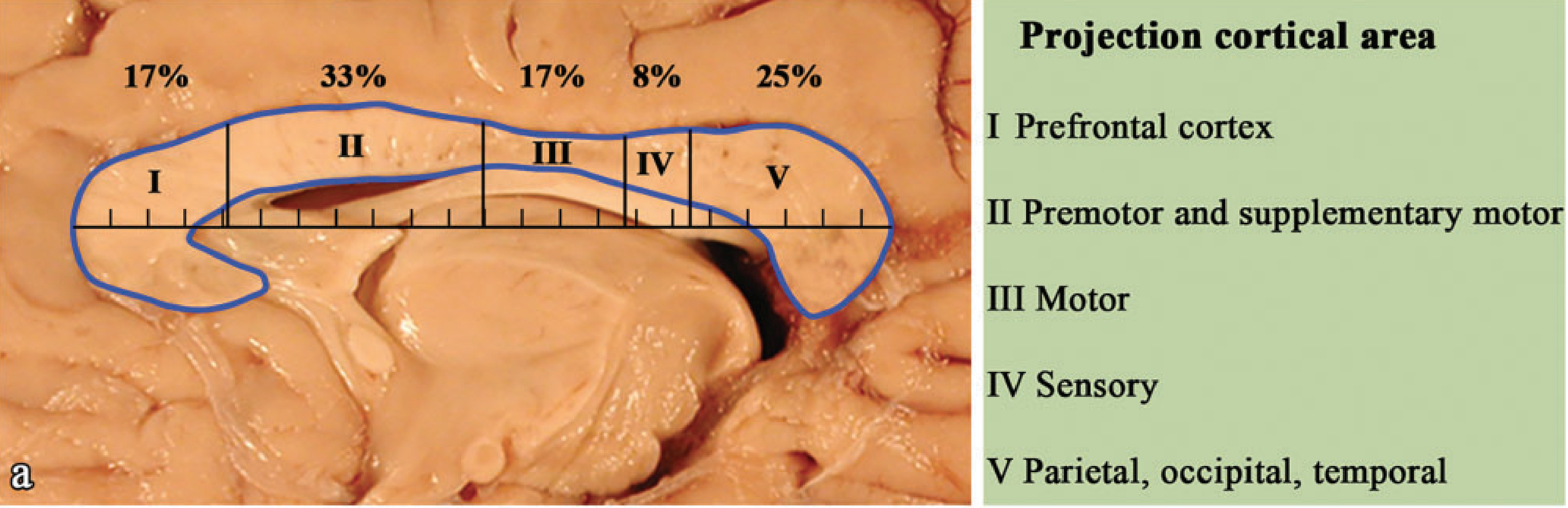
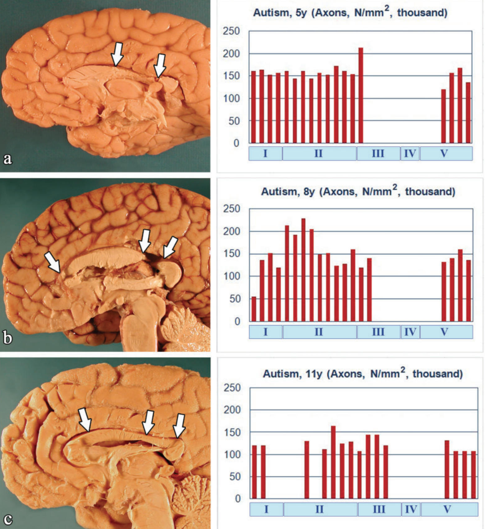
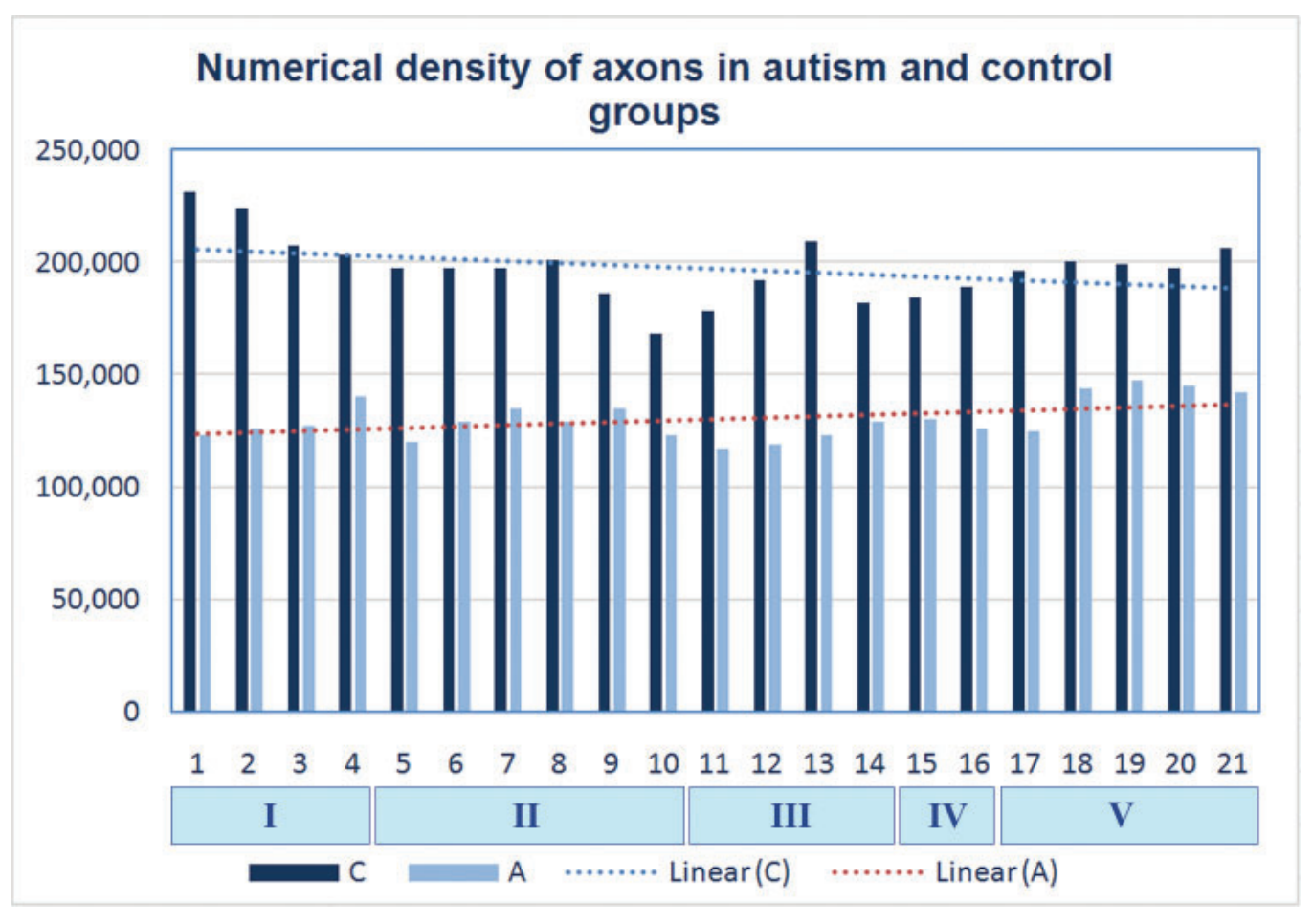
Wegiel and his group 3 studied the corpus callosum of 11 people with autism, ages 4 to 60, of whom eight were men and three were women, and 11 other controls, ages 4 to 52, of whom seven were men and four were women. In the brains of three autistic people, hypogenesis of the corpus callosum resulted in partial or total loss of the interhemispheric axonal connections in segments III-V of the corpus callosum. In these cases, the combination of a focal genesis and a uniform axonal deficit generated a reduction in the areas of the corpus callosum by 37%, the number of axons by 62% and the numerical density of axons by 39%. Temur and his group 4 have confirmed in autopsy material that the area and volume of the corpus callosum in children with ASD is less than in controls. They have also seen that there is microstructural damage that seems to correspond to a reduction in the myelinization of the nerve tracts, altered integrity, reduction in axonal density and changes in the organization of the white substance.
Callus abnormalities are not uncommon. The agenesis of the corpus callosum, being born without this connection between the two hemispheres, or hypogenesis, which is minor at birth, appears in more than fifty congenital syndromes that are associated with brain malformations. In the United States, various abnormalities of corpus callosum development affect between 0.7% and 5.3% of the general population. The prevalence of the most striking, the agenesis of the corpus callosum, the total disappearance of this important commissure, is according to various studies from 1 per thousand to 1 in four thousand 5. Complete or partial agenesis is often associated with intellectual deficits and a wide range of cognitive, behavioral and neurological consequences. Many individuals with corpus callosum agenesis have been diagnosed, often without knowledge of this anatomical abnormality, with cognitive impairment, attention deficit and/or autism spectrum disorder.
Agenesis of the corpus callosum and autism may coincide, but not always. Unusual social interactions have been observed in almost half (40%) of children with corpus callosum agenesis, but the most common differences are emotional immaturity, lack of introspection, altered social competence, deficits in social judgment and planning, and poor communication of emotions. On the other hand, there are differences between people with autism and those with corpus callosum agenesis, including an onset of social deficit at age 2-3 in people with autism and at age six in children with corpus callosum agenesis. Repetitive behaviors and restricted interests are less common in children with corpus callosum agenesis than in children with autism. People with corpus callosum agenesis show deficits in communication and social interaction, which overlap with the diagnostic criteria for autism. 8.5% of children with corpus callosum agenesis have a diagnosis of autism, while the proportion is 1% in their siblings.
The conclusion of these studies is that the corpus callosum appears to be one of the key brain tracts in explaining some behaviors commonly associated with autism.
References
- Chung MK, Dalton KM, Alexander AL, Davidson RJ (2004) Less white matter concentration in autism: A 2D voxel-based morphometry. Neuroimage 23: 242–251. doi: 10.1016/j.neuroimage.2004.04.037. ↩
- Hardan AY, Minshew NJ, Keshavan MS (2000). Corpus callosum size in autism. Neurology 55(7), 1033–1036. doi:10.1212/wnl.55.7.1033 ↩
- Wegiel J, Flory M, Kaczmarski W, Brown WT, Chadman K, Wisniewski T, Nowicki K, Kuchna I, Ma SY, Wegiel J (2017) Partial agenesis and hypoplasia of the corpus callosum in idiopathic autism. J Neuropathol Exp Neurol 76(3):225-237. doi: 10.1093/jnen/nlx003. ↩
- Temur HO, Yurtsever I, Yesil G, Sharifov R, Yilmaz FT, Dundar TT, Alkan A (2019) Correlation between DTI findings and volume of corpus callosum in children with autism. Curr Med Imaging Rev 15(9): 895-899. doi: 10.2174/1573405614666181005114315. ↩
- Paul LK, Brown WS, Adolphs R, Tyszka JM, Richards LJ, Mukherjee P, Sherr EH (2007) Agenesis of the corpus callosum: genetic, developmental and functional aspects of connectivity. Nat Rev Neurosci 8(4): 287-299. doi: 10.1038/nrn2107. ↩
1 comment
[…] Autismoaren ezaugarri fisiko esanguratsuenetarikoa izan daiteke garunaren bi hemisferioak batzen dituen gorputz kailukararean berezitasunak. José Ramón Alonsoren Corpus callosum and autism […]