Expansion microscopy: a new technique to see inside microbes
Expansion microscopy: a new technique to see inside microbes
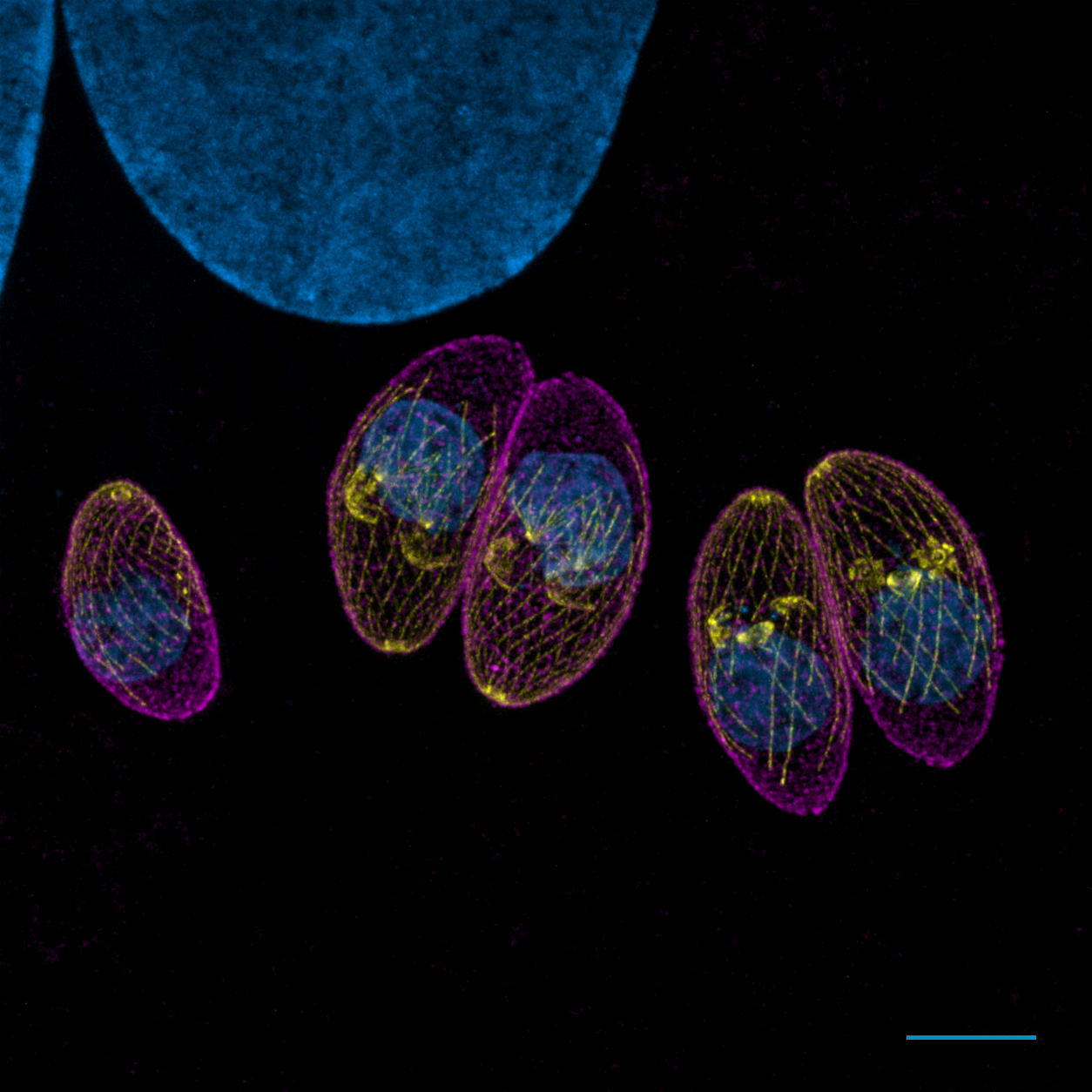
How can scientists see the intricate details inside cells far too small for regular light microscopes? A powerful technique called expansion microscopy 1 is revolutionizing how researchers study tiny organisms from plankton to developing embryos.
Making the invisible visible
Expansion microscopy works by doing the opposite to what we had been doing until now: instead of using more powerful lenses to see smaller structures, scientists physically expand the samples themselves. To do so, they embed cells or tissues in a clear gel that swells when it absorbs water like a sponge, magnifying the sample up to 16 times its original size!
Remarkably, the internal structures remain intact during this process and expand proportionally, allowing researchers to see details that would normally require expensive, specialized equipment. The technique has proven especially valuable for studying microbial eukaryotes—tiny organisms notoriously difficult to examine as many cannot be genetically modified to make their structures glow under microscopes and/or their tough cell walls prevent traditional staining methods from working.
Exploring ocean life at unprecedented scale
One major application involves studying plankton, the microscopic organisms floating in the ocean that produce most of our planet’s oxygen and are the basis of marine food chains. Using this new technique, scientists observed hundreds of planktonic species, revealing diverse patterns of organisation of the cytoskeleton—the internal scaffolding that helps cells maintain their shape, divide, and move.
By analyzing these structures across many species, researchers could make predictions about how cellular organization evolved over millions of years. 2 This will allow us to develop an encyclopedia of planktonic cellular architecture that links physical structure to evolution.
The technique also allows studying diatoms, microscopic algae with glass-like silica cell walls and extremely difficult to study conventionally. However, with expansion microscopy researchers could overcome 3 previous limitations and examine their internal structures.
Beyond microscopic organisms, researchers are applying expansion microscopy to understand tissue development. Scientists studying limb formation in mouse embryos used it to observe how cells orient themselves during development, preserving three-dimensional spatial information that would be lost with traditional methods.
Unlike electron microscopy requiring expensive specialized equipment, expansion microscopy can be performed with standard laboratory microscopes, making it accessible to more research teams worldwide. This makes the technique a go-to resource for looking into the intricate structures of living organisms as they are, without the distortion associated with most other methods of fixating and labelling samples for imaging.
References
- Wassie, A.T., Zhao, Y. & Boyden, E.S. (2019) Expansion microscopy: principles and uses in biological research. Nat Methods doi: 10.1038/s41592-018-0219-4 ↩
- Mikus F, Rubio Ramos A, Shah H. et al (2025) Charting the landscape of cytoskeletal diversity in microbial eukaryotes
Cell doi: 10.1016/j.cell.2025.09.027 ↩ - Flori S, Mikus F, Flaum E. et al (2025) Diatom ultrastructural diversity across controlled and natural environments Current Biology doi: 10.1016/j.cub.2025.10.024 ↩