Hibernation and white-nose syndrome
Hibernation and white-nose syndrome
Hibernation is a state of inactivity and metabolic depression used by some mammals to survive a cold winter. It may last several days, weeks, or months depending on the species, environment temperature, season, and individual’s body condition. Usually, hibernating animals survive the energetic bottleneck of the winter by building stores of body fat in late summer and early autumn and by conserving energy through extended periods of low activity. Small hibernating animals have a torpor-arousal cycle, with torpor bouts persisting for several days or weeks whereas the euthermic arousal bouts last less than a day. During torpor, many basic organic activities such as body temperature, heart contraction, mitochondrial respiration, oxygen consumption, and blood flow are reduced to a minimum that would be lethal to most homeotherms. Heart rates wind down to a mere 10 beats a minute and body temperature become near to environment temperature.
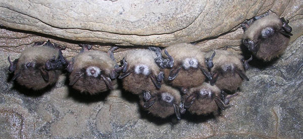
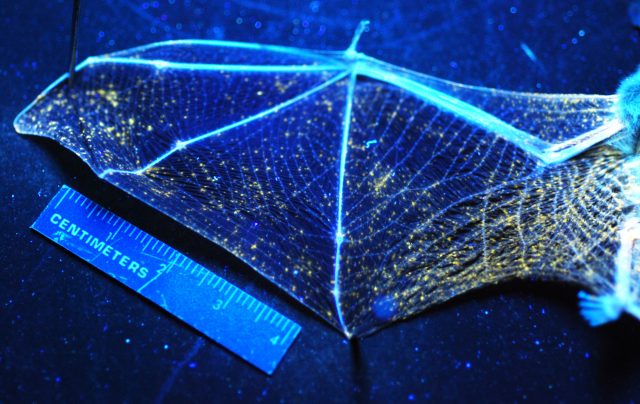
Bats account for a quarter of mammalian species. Not all bats hibernate, but those that do usually live at high latitudes where insect prey becomes scarce during cold months. Unfortunately, white-nose syndrome, a fatal fungal affliction is devastating hibernating bats across the eastern half of North America. The disease is caused by the psychrophilic fungus Pseudogymnoascus destructans, an ascomycete fungal pathogen found in Europe, Asia and North America. Since it emerged in New York State in 2007, millions of bats have died and mortality ranges from 95% to 100% of infected animals. By contrast, there has been no significant mortality in Europe, despite wide distribution and high prevalence of white-nose syndrome in many European bat hibernacula. The fungus is invasive and damages the dermal tissue on the wings of bats during hibernation, forming characteristic long-lasting erosions that cause inflammation and lead to fluid and electrolyte loss across the damaged dermal tissue leading to a cascade of physiological alterations. Although the mechanisms are nowadays unknown, the main problem seem to be that sick bats behave abnormally, disrupt their hibernation by increased and abnormal arousals from torpor and go on daytime flights during the winter, a process that leads to premature depletion of lipid reserves, leaving them weak and emaciated. The average torpor bout length in unaffected little brown myotis is circa 13 days whereas the little brown myotis dying of the infection have decreased torpor bout length to approximately 8 days. 1 Thus, it is important what happens during torpor-arousal cycles and what signals take the animals out from hibernation.
The question of why hibernators may experience the periodic arousals (returns to normal body temperature) has intrigued investigators for decades and there are many hypotheses on the subject. One favored idea is that hibernators build a ‘sleep debt’ during hibernation, and so must occasionally warm up in order to sleep and do the required brain maintenance. Another theory states that the brief periods of high body temperature during hibernation are used by the animal to restore its available energy sources. Yet another hypothesis proposes that the returns to high body temperature allow mammals to initiate an immune response. These cycles have features similar to a repeated process of ischemia and reperfusion. During this latter phase, the quick restoration of blood circulation accompanied by an increased mitochondrial respiration and oxygen usage may result in elevated generation of reactive oxygen species (e.g. H2O2 and ROO.) and reactive nitrogen species (e.g., NO and ONOO.-). These chemical species produce oxidative damage and are involved in ageing and several diseases including Parkinson’s, Alzheimer´s, diabetes and stroke.
Brain is the most hypoxia-sensitive organ and is highly vulnerable to oxidative damage. A research group from East China Normal University in Shanghai has studied the antioxidant defenses in the brain of bats during hibernation 2. They found that the total level of reactive oxygen species and reactive nitrogen species in the brain of each of the two distantly related hibernating bats Myotis ricketti and Rhinolophus ferrumequinum at arousal was lower than that at torpid or active state. This observation suggests that bats maintain a basal level of reactive oxygen species and reactive nitrogen species that does no damage the brain during hibernation. Hibernating animals have an antioxidant defense system that uses antioxidant proteins, low molecular weight antioxidants (glutathione, urate, and ascorbate) and malondialdehyde.
The system is probably even more complex. Other hibernating animals such as the Arctic ground squirrels, that may exhibit abdominal temperatures as low as -2,9 ºC, do not show detrimental effects of global cerebral ischemia in vivo, an effect that is also evident in neuronal progenitor cells. Whereas human neuronal progenitor cells are damaged by ischemia/reperfusion injury, similar cells from the hibernating squirrels resist cell death and show evidence of proliferation in response to hypoxia 3.
Nevertheless, the key item seems to be arousal. There is evidence of surviving populations of little brown myotis bats (Myotis lucifugus) close to where the fungus was first detected nearly ten years ago in New York State. Despite infection with Pseudogymnoascus destructans, the causative fungal agent, the remnant population displayed less frequent arousals from torpor and lower torpid body temperatures than bats that died from white-nose syndrome during the peak of mortality. These animals do show the fungal pathogen on their skin but they do not experience the increase in periodic arousals from hibernation typified by bats dying from the syndrome.
References
- Lilley TM, Johnson JS, Ruokolainen L, Rogers EJ, Wilson CA, Schell SM, Field KA, Reeder DM (2016) White-nose syndrome survivors do not exhibit frequent arousals associated with Pseudogymnoascus destructans infection. Front Zool 13: 12. ↩
- Yin Q, Ge H, Liao CC, Liu D, Zhang S, Pan YH (2016) Antioxidant Defenses in the Brains of Bats during Hibernation. PLoS One 11(3): e0152135. ↩
- Drew KL, Wells M, McGee R, Ross AP, Kelleher-Andersson J (2016) Arctic ground squirrel neuronal progenitor cells resist oxygen and glucose deprivation-induced death. World J Biol Chem 7(1): 168-177. ↩