Predicting the shape of a fat-lengthening enzyme to guide drug design
Predicting the shape of a fat-lengthening enzyme to guide drug design
Fats do far more than store energy. They form the membranes that enclose every cell and act as chemical signals. Cells build and reshape them with enzymes, and one of these, ELOVL6, lengthens fatty acids by two carbon atoms. Embedded in the membrane of the endoplasmic reticulum, an internal network where many lipids are made, and most abundant in liver and fat tissue, it converts 16-carbon fatty acids into 18-carbon ones, such as palmitic acid into stearic acid. The change looks small, yet chain length alters how fats behave in cells.
Medical interest began in the early 2000s. ELOVL6 was characterized in mouse in 2002 as a gene switched on by a master regulator of fat production. In 2007, mice lacking it still became obese on a fatty diet but were largely protected from insulin resistance, the disturbance behind type 2 diabetes. Since then the enzyme has been linked to fatty liver disease and atherosclerosis, and more recently to certain cancers and to the failure of nerve-insulating myelin to regrow in multiple sclerosis. These are associations, not proof that blocking the enzyme would treat any of these conditions, but they make it an attractive drug target.
Designing a drug usually requires the three-dimensional shape of its target, because most drugs work by fitting into a pocket on a protein. Since the first protein structure, myoglobin, was solved in the late 1950s, X-ray crystallography has supplied most such shapes, but membrane-embedded proteins like ELOVL6 are notoriously hard to crystallize. Of the seven human ELOVL enzymes, only ELOVL7 has been solved, in 2021. It revealed a narrow tunnel running into the membrane that cradles the fatty acid chain, with the reaction site deep inside. Potent ELOVL6 inhibitors were reported around 2009, and some clearly preferred ELOVL6 over its relatives, yet none has reached clinical use, and how they bind has remained unknown.
A recent computational study 1 tackled this gap in layers. It first built a model of ELOVL6 with AlphaFold, the artificial intelligence system that since 2020 has predicted protein shapes from amino acid sequences with accuracy approaching experiments, and which earned a share of the 2024 Nobel Prize in Chemistry. A newer version, AlphaFold3, produced nearly identical shapes, and the model resembled ELOVL7’s measured structure, which is slightly longer to hold longer chains.
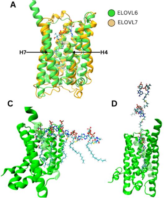
Next, simulations that treat atoms as particles following physical laws explored how a fatty acid attached to coenzyme A, a carrier molecule, might reach the reaction site. Two routes were compared: inserting the fatty acid chain first, tail-end down the narrow tunnel, or sliding the whole molecule in sideways through a gap between two spiral-shaped protein segments, called helices H4 and H7. Sideways entry was far cheaper in energy, by about 13 kilocalories per mole, and in every trial helix H4 shifted to widen the gap. Because these simulations took place in water rather than a membrane, this remains a prediction, not an observation.
The study then asked where inhibitors bind. Three candidate pockets were tested: the side gap, an outer site that might alter the enzyme’s shape from a distance, and the reaction site itself. For two well-studied inhibitors, calculated binding strengths and a virtual screening test favored the reaction site. In that test, about 3,400 compounds, mostly decoys assumed not to be active, were ranked to see whether known inhibitors rose to the top. Calculated strengths at the reaction site roughly matched laboratory values, whereas the other pockets fell well short. Even so, the raw AlphaFold pocket ranked inhibitors only modestly better than chance and looked too rigid.
Refining it meant generating hundreds of candidate arrangements of each inhibitor, discarding unstable ones, and keeping those whose predicted potency differences among related compounds matched laboratory measurements. The refined models picked out known inhibitors far more reliably, including those from a chemical family different from the one used to build them. A glutamine at position 202 formed a stable hydrogen bond, a weak attraction involving hydrogen, with both inhibitors, and requiring this contact improved the screening further.
The study also probed why the inhibitors favor ELOVL6. Swapping amino acids lining the pocket to mimic related enzymes reproduced many, though not all, of the measured differences. A second set of simulations, which tracked how flexible each pocket is, added a subtler explanation: ELOVL6 and its closest relative, ELOVL3, often open a pocket large enough for the bulkier inhibitor, whereas ELOVL1, ELOVL2 and ELOVL5 rarely do, so binding would cost them an energetic penalty. Selectivity therefore seems to depend on both amino acid differences and flexibility.
Important caveats remain. ELOVL6’s structure has still not been determined experimentally, every result here is computed, and some simulations simplified the membrane. Strong binding also does not make a compound a safe or effective medicine, as the earlier inhibitors show. Still, the models offer a testable framework that may help scientists screen large compound libraries and predict how chemical changes affect binding, narrowing the candidates worth testing in the laboratory.
Author: César Tomé López is a science writer and the editor of Mapping Ignorance
Disclaimer: Parts of this article may have been copied verbatim or almost verbatim from the referenced research paper/s.
References
- M. G. Ibarluzea, R. Ramis, M. Fuentetaja, F. J. Gil-Bea, G. Gerenu, A. Lopez De Munain, J. M. Aizpurua, J. I. Miranda, A. Bergara, and A. Leonardo (2026) Computational Analysis of ELOVL6 Structure and Inhibition for Rational Drug Design J. Chem Inf. Model. doi: 10.1021/acs.jcim.6c00336 ↩